Microphysiological Systems: Advancing Drug Development and Personalized Medicine
Foundations of Microphysiological Systems
Microphysiological systems (MPS) represent a culmination of advances in several scientific domains aimed at recreating human physiology outside the body. They harness the principles of systems biology, stem cell biology, and regenerative medicine to replicate the complex interplay of tissues and organs.
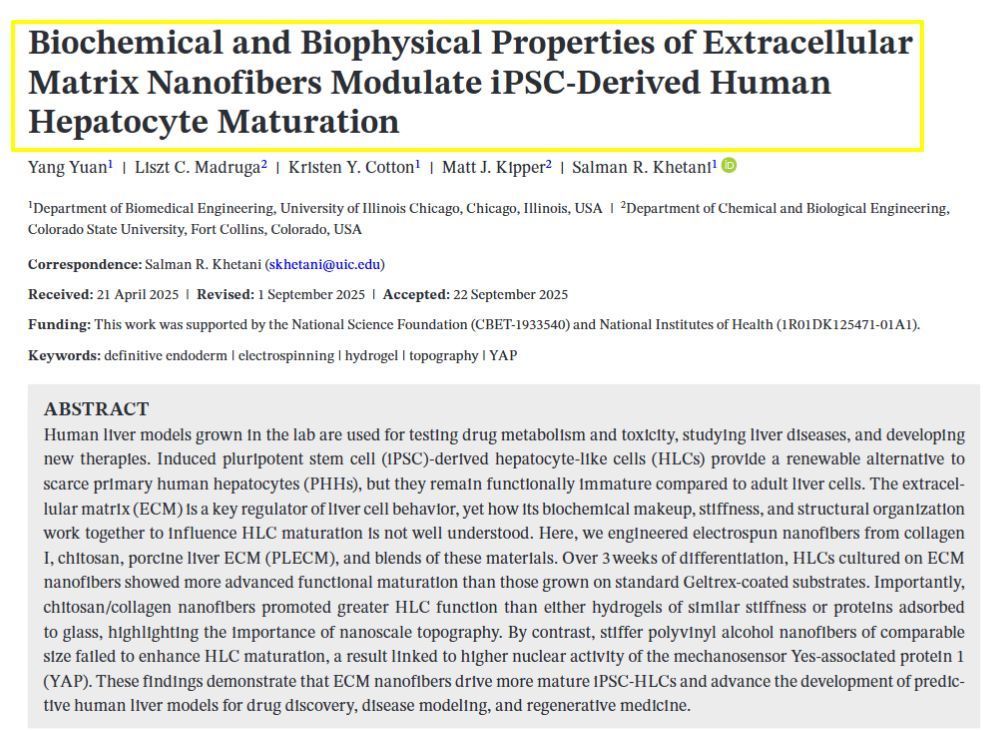
Biomaterials play a crucial role in MPS, providing scaffolds that mimic the three-dimensional architecture of biological tissue. This structural complexity allows for more physiologically relevant cell cultures than traditional two-dimensional petri dishes. MPS technology often incorporates organs-on-chips or tissue chips, where microfluidics create an environment that simulates blood flow and other mechanical forces experienced by organs.
The integration of pluripotent and induced pluripotent stem cells facilitates the generation of diverse cell types, offering a window into human physiology and disease mechanisms. These cells possess the plasticity required for creating tissue constructs that resemble real organ functionality.
In drug development, MPS provide an invaluable platform for efficacy and toxicology testing, reducing the reliance on animal models and improving the predictability of human responses to new compounds. Through the combination of tissues and their interactions, MPS strive to replicate organ culture within a controlled setting that closely emulates living systems.
The interplay of these components within MPS mirrors organ-level functions, making them a potent tool in the understanding and manipulation of human physiology for medical and research applications.
Applications in Drug Discovery and Development
Microphysiological systems (MPS) are revolutionizing drug discovery and development by offering improved models for toxicology, disease modeling, and pharmacology. These systems enhance the predictive accuracy of drug responses and safety profiles, thereby potentially reducing the attrition of drug candidates.
Toxicology and Drug Safety
MPS provide a dynamic platform for toxicology studies by simulating human tissue and organ responses to new compounds. Drug safety assessments benefit from these systems as they allow for the evaluation of drug exposure and toxicity with higher precision than traditional models. This capability facilitates early-stage risk assessment and informs safety profiles, in hopes of decreasing clinical trial failure rates due to adverse effects.
Disease Modeling and Organoids
In the realm of disease modeling, MPS can be designed to replicate disease states within organ-specific contexts. Organoids—miniaturized, simplified versions of organs—play a pivotal role here. They enable researchers to observe disease progression and organ functions in a controlled environment, which can lead to a deeper understanding of disease mechanisms and more effective drug discovery initiatives, moving towards personalized medicine.
Pharmacology and Drug Screening
Pharmacology studies benefit from MPS's capacity to model the complex interactions between drugs and biological systems. They have become an integral tool in drug screening processes by providing insights into the efficacy and mechanisms of action of new compounds. Utilizing these in vitro systems, researchers can rapidly assess the therapeutic potential of multiple drug candidates, thus accelerating the drug development lifecycle and improving clinical outcomes.
Technological Advancements and Challenges
The integration of bioengineering and microfluidic technologies heralds a progressive era for in vitro models, yet the road ahead is marked by both promise and considerable obstacles.
Microfluidic Technologies and Organs-on-a-Chip
Microfluidic technologies form the backbone of Organs-on-a-Chip (OoC) systems, harnessing miniature channels to replicate the fluid flow and cellular environments of human organs. These bioengineered devices have proven instrumental in studying drug metabolism, facilitating drug discovery, and providing insights into environmental toxicology. Multi-organ MPS, or Physiome-on-a-Chip, represents a cutting-edge implementation, aiming to mimic the complex physiology of human organ interactions.
Advances in the field:
- National Institutes of Health and National Academies of Sciences, Engineering, and Medicine have recognized the significance of this technology.
- Progress in creating human and animal MPS banks offers a portfolio of tools for researching systemic responses to substances.
Challenges in Implementation
However, significant challenges obstruct broader adoption in the pharmaceutical industry and other fields. Key among these is the reproducibility of results, as minor deviations in the chip manufacturing or operating processes can lead to drastically different outcomes.
Limitations:
- Universal standards for construction and operation are still lacking.
- Complexity in replicating the exact physiological conditions of human tissue remains a hurdle.
- Necessity for areas of needed improvement such as long-term stability and integration into existing workflows.
By confronting these challenges, developers and researchers aim to refine microphysiological systems into reliable platforms for hazard identification and broader applications in human health and disease modeling.


