Grants Awarded to the MTM Laboratory
2024
- The MTM Lab has been awarded an NIDDK R01 (National Institute of Diabetes and Digestive and Kidney Diseases) grant to develop a novel microfluidic approach to elucidate the effects of soluble factor gradients, individually and in controlled combinations, on zonated functions in primary liver cells from rodents and humans towards determining species-specific effects. Ultimately, our novel devices can be used to investigate the mechanisms underlying liver zonation, chemical-induced zonated hepatotoxicity, and how zonation is perturbed in liver diseases, such as non-alcoholic fatty liver disease and hepatocellular carcinoma.
- The MTM Lab has been awarded a NIEHS (National Institute of Environmental Health Sciences) grant to develop a high throughput system to test placental cell invasion using a 3D placental microtissue coupled with hepatic liver biotransformation. This first-of-its-kind hepatic-placenta organ-tandem on a chip will simulate the liver metabolism that chemicals undergo in vivo prior to reaching the placental bed. This state-of-the-art in vitro platform will be the first step towards incorporating organism-level organization into reproductive risk assessment using a non-animal-based approach.
- The MTM Lab has been awarded a NIEHS (National Institute of Environmental Health Sciences) grant to develop a human gut-liver platform with microbiome interactions for in vitro toxicology. These first-of-its-kind scalable human gut-liver models will be developed for in vitro applications, such as compound screening and disease modeling, and be used to elucidate the effects of reciprocal tissue crosstalk on cell phenotype modulation.
2023
- The MTM Lab has been awarded a NIDDK (National Institute of Diabetes and Digestive and Kidney Diseases) grant to analyze the synergistic effects of extracellular matrix composition and stiffness, multicellular interactions, and soluble triggers of NAFLD in cellular phenotypic alterations, which could aid the development of novel drug therapies for this disease.
- The MTM Lab has been awarded a NIAAA (National Institute on Alcohol Abuse and Alcoholism) grant to develop a first-of-its-kind organotypic mouse liver model and investigate the effects of alcohol on multiple liver cell types in this model with comparisons to an in vivo mouse model of ALD that recapitulates several key features of human ALD. This platform can aid in understanding the molecular mechanisms underlying alcohol-associated liver disease.
2021
- The MTM lab, in collaboration with the Shah lab, has been awarded an NIH R21 grant to develop design rules for the bioprinting of 3D human liver tissue surrogates that have three key integrated compartments as in the human body: hepatic, vascular, and biliary. This work will advance liver co-culture systems and biomaterials most suitable for 3D printing of the liver.
- The MTM lab, in collaboration with the Rehman, Merrill, and Dai labs, has been awarded an NSF grant to develop synergistic microenvironmental, computational, and genetic approaches for reproducibly biofabricating functionally mature liver organoids for drug testing.
- The MTM lab, in collaboration with the Pajcini Lab, has been awarded an NIH R01 grant to develop human liver models for the study and expansion of hematopoietic stem cells.
2020
- The MTM lab is collaborating and receiving funding from an NIH R01 grant (PI: Dr. Dawood Darbar, Co-I: Dr. Salman Khetani) focused on determining the genetic determinants of atrial fibrillation and help develop better therapies (with clinical trials) for patients suffering from this most common sustained arrhythmia.
- The MTM lab has been awarded a National Heart, Lung, and Blood Institute (NHLBI) R01 grant to develop, in close collaboration with Dr. Dawood Darbar , Chief of the Division of Cardiology at the University of Illinois Medical School, in vitro models of atrial fibrillation (the most common sustained arrhythmia) using patient-derived induced pluripotent stem cells that can be used to elucidate underlying genetic mechanisms of this disease and for novel drug discovery.
2019
- The MTM lab has been awarded a National Institute of Environmental Health Sciences (NIEHS) R21 grant to explore, in collaboration with the Underhill lab at UIUC, the role of key biochemical and biophysical microenvironmental cues on the functions of induced pluripotent stem cell-derived human hepatocyte-like cells +/- co-culture with liver sinusoidal endothelial cells.
- The MTM lab has been awarded a National Science Foundation grant to develop, in collaboration with Dr. Matthew Kipper's group at Colorado State University, nanostructured 3-dimensional scaffolds that can be used to deliver physiological biochemical and biophysical cues to stem cell-derived human liver cells to further mature their functions in vitro for drug screening with eventual utility for regenerative medicine.
- The MTM lab has been awarded an NIH R21 grant to develop a scalable 3D human liver platform for investigating the pathogenesis of hepatitis B viral infection.
- The MTM lab has received funding from the Center for Advanced Design and Manufacturing of Integrated Microfluidics (CADMIM) to develop, in collaboration with the Hui lab at the University of California at Irvine, a cell patterning method using DNA template-based assembly.
2018
- The MTM lab has been awarded an NIH R01 grant, in collaboration with the Underhill Lab at UIUC, to explore molecular mechanisms and develop a drug screening platform for non-alcoholic fatty liver disease.
2017
- The MTM lab has been awarded an NSF grant to develop a high-throughput microliver platform for drug toxicity screening in collaboration with Dr. David Wood's Living Devices Lab at the University of Minnesota.
- The MTM lab is awarded an NIH R21 grant to explore, in collaboration with Dr. David Wood's Living Devices Lab at the University of Minnesota, the role of key microenvironmental factors on the functional maturation of induced pluripotent stem cell-derived human liver cells using a novel 3D microgel platform.

Recent advances in biofabrication are revolutionizing liver tissue engineering by enabling precise spatial patterning of liver cells to mimic the organ’s complex architecture. Techniques like 3D bioprinting, microfluidics, and self-assembled cell aggregates help recreate critical features such as metabolic zones, cell polarity, and vascular networks. These engineered liver models improve drug testing, disease research, and hold promise for regenerative therapies. Despite challenges in scaling and standardization, integrating multiple fabrication methods and emerging technologies like machine learning are driving progress. Ultimately, these innovations bring us closer to creating functional liver tissues for clinical and pharmaceutical applications. See full article here.
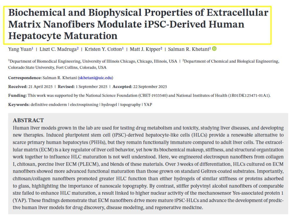
𝗦𝗶𝗺𝗽𝗹𝗲 𝗦𝘂𝗺𝗺𝗮𝗿𝘆: This study explores how we can improve lab-grown liver cells for medical research and drug testing. The MTMLab team works with induced pluripotent stem cells (iPSCs) - special cells that can be transformed into liver-like cells - because real human liver cells are hard to obtain. However, these lab-grown liver cells don't function as well as mature adult liver cells. The research team discovered that the surface environment where these cells grow is crucial for their development. We created tiny fiber scaffolds made from different materials like collagen, decellularized livers, and chitosan that mimic the natural structure around liver cells. When liver cells were grown on these specially designed nanofibers for three weeks, they displayed higher function compared to cells grown on standard surfaces. Our key finding was that both the material composition and the nanoscale fiber structure were important - stiffer synthetic fibers or softer materials without the appropriate topography or composition prevented proper cell maturation. This research helps create better lab models of human liver tissue that can be used for testing new drugs and studying liver diseases more effectively.
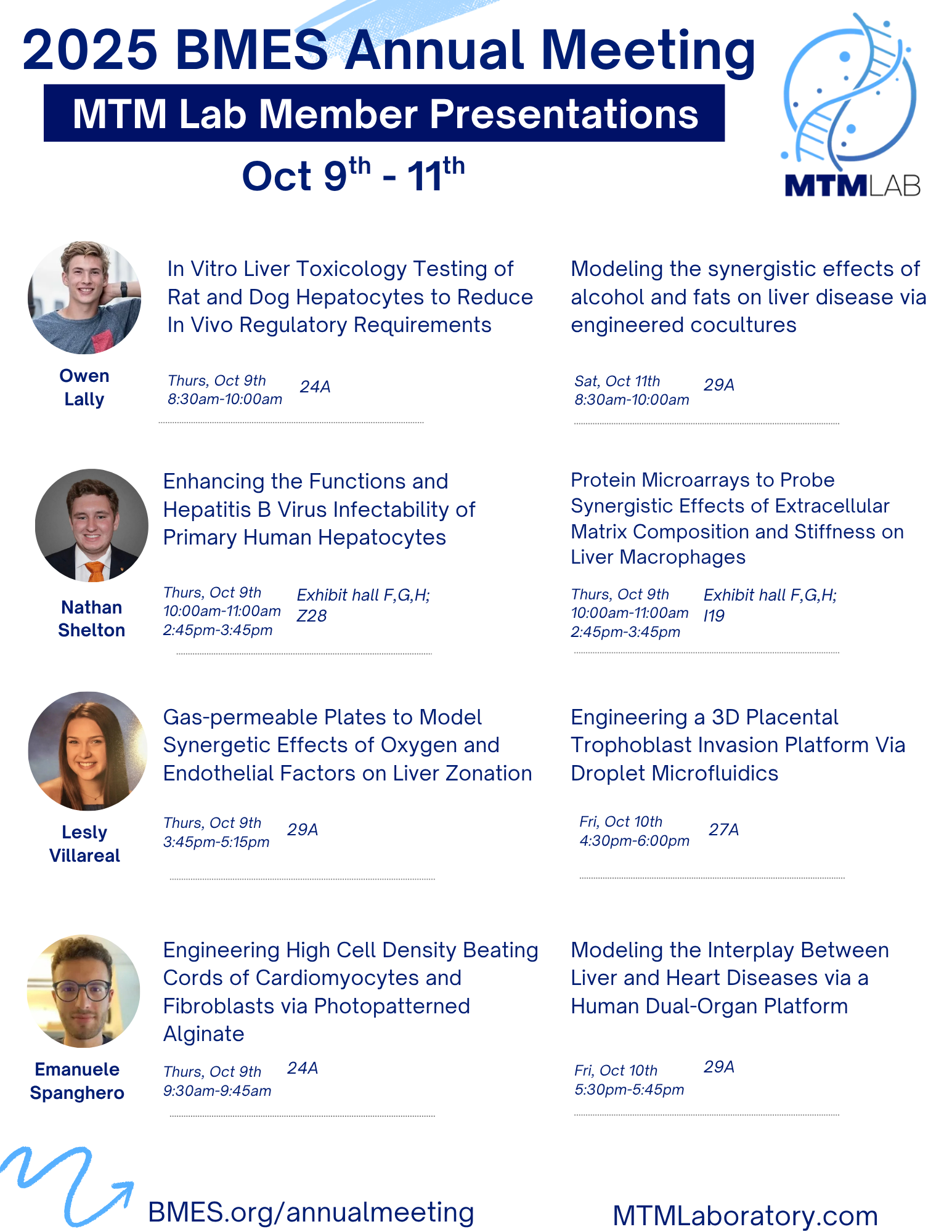
Owen Lally Modeling the synergistic effects of alcohol and fats on liver disease via engineered cocultures In Vitro Liver Toxicology Testing of Rat and Dog Hepatocytes to Reduce In Vivo Regulatory Requirements Nathan Shelton Enhancing the Functions and Hepatitis B Virus Infectability of Primary Human Hepatocytes Protein Microarrays to Probe Synergistic Effects of Extracellular Matrix Composition and Stiffness on Liver Macrophages Lesly Villarreal Engineering a 3D Placental Trophoblast Invasion Platform Via Droplet Microfluidics Gas-permeable Plates to Model Synergetic Effects of Oxygen and Endothelial Factors on Liver Zonation Emanuele Spanghero Modeling the Interplay Between Liver and Heart Diseases via a Human Dual-Organ Platform Engineering High Cell Density Beating Cords of Cardiomyocytes and Fibroblasts via Photopatterned Alginate