MTM Lab Students Present at Major Conferences & Symposia

- Students in the MTM lab presented 12 abstracts at the 2023 Annual Meeting of the Biomedical Engineering Society in Seattle, WA.
- Students in the MTM lab presented 11 abstracts in the form of 7 platform talks and 4 posters at the 2021 Annual Meeting of the Biomedical Engineering Society in Orlando, FL.
- Regeant Panday, a PhD student in the MTM lab, presented his work with 3D human liver tissues (poster) at the MicroTAS 2020 conference.
- The MTM lab presented 6 accepted abstracts (2 talks and 3 posters) at the Annual Meeting of the Biomedical Engineering Society.
- The MTM lab presented 8 abstracts, one as an oral presentation and seven as poster presentations at The Second Annual UIC Bioengineering Research Symposium. Congratulations to Grace Brown, Hardik Dabas, Demi Ibrahim, David Kukla, Jennifer Liu, Chase Monckton, Regeant Panday, and Yang Yuan for these presentations.
- The MTM lab presented 3 posters at the biannual meeting of the Center for Advanced Design and Manufacturing of Integrated Microfluidics (CADMIM) in Irvine, CA. Congratulations to Jennifer Liu, Grace Brown, and David Kukla for these presentations.
- The MTM lab presented 8 abstracts at the annual meeting of the Biomedical Engineering Society (BMES), incluidng 3 oral presentations and 5 poster presentations. Congratulations to Grace Brown, David Kukla, Jennifer Liu, and Chase Monckton for these presentations.
- Dr. Khetani presented MTM lab's research on a microfluidic human liver model at the annual meeting of the Biomedical Engineering Society (BMES) in Phoenix, AZ.
- David Kukla , Matt Davidson and Dr. Khetani presented MTM research in the form of oral talks and poster presentations at the annual meeting of the Biomedical Engineering Society (BMES) in Minneapolis, MN.
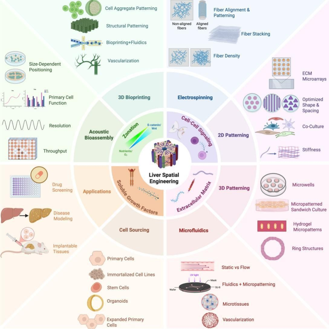
Recent advances in biofabrication are revolutionizing liver tissue engineering by enabling precise spatial patterning of liver cells to mimic the organ’s complex architecture. Techniques like 3D bioprinting, microfluidics, and self-assembled cell aggregates help recreate critical features such as metabolic zones, cell polarity, and vascular networks. These engineered liver models improve drug testing, disease research, and hold promise for regenerative therapies. Despite challenges in scaling and standardization, integrating multiple fabrication methods and emerging technologies like machine learning are driving progress. Ultimately, these innovations bring us closer to creating functional liver tissues for clinical and pharmaceutical applications. See full article here.
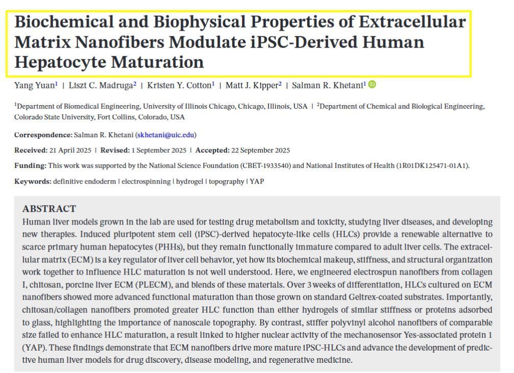
𝗦𝗶𝗺𝗽𝗹𝗲 𝗦𝘂𝗺𝗺𝗮𝗿𝘆: This study explores how we can improve lab-grown liver cells for medical research and drug testing. The MTMLab team works with induced pluripotent stem cells (iPSCs) - special cells that can be transformed into liver-like cells - because real human liver cells are hard to obtain. However, these lab-grown liver cells don't function as well as mature adult liver cells. The research team discovered that the surface environment where these cells grow is crucial for their development. We created tiny fiber scaffolds made from different materials like collagen, decellularized livers, and chitosan that mimic the natural structure around liver cells. When liver cells were grown on these specially designed nanofibers for three weeks, they displayed higher function compared to cells grown on standard surfaces. Our key finding was that both the material composition and the nanoscale fiber structure were important - stiffer synthetic fibers or softer materials without the appropriate topography or composition prevented proper cell maturation. This research helps create better lab models of human liver tissue that can be used for testing new drugs and studying liver diseases more effectively.
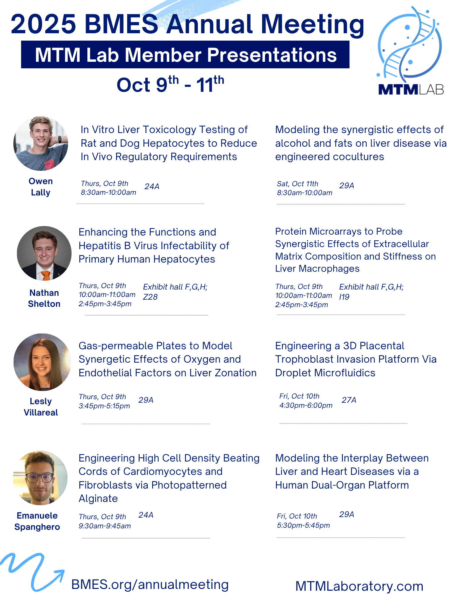
Owen Lally Modeling the synergistic effects of alcohol and fats on liver disease via engineered cocultures In Vitro Liver Toxicology Testing of Rat and Dog Hepatocytes to Reduce In Vivo Regulatory Requirements Nathan Shelton Enhancing the Functions and Hepatitis B Virus Infectability of Primary Human Hepatocytes Protein Microarrays to Probe Synergistic Effects of Extracellular Matrix Composition and Stiffness on Liver Macrophages Lesly Villarreal Engineering a 3D Placental Trophoblast Invasion Platform Via Droplet Microfluidics Gas-permeable Plates to Model Synergetic Effects of Oxygen and Endothelial Factors on Liver Zonation Emanuele Spanghero Modeling the Interplay Between Liver and Heart Diseases via a Human Dual-Organ Platform Engineering High Cell Density Beating Cords of Cardiomyocytes and Fibroblasts via Photopatterned Alginate