What Are iPSC-derived Hepatocytes? Why Are They Important?

iPSC-derived hepatocytes are liver cells created from induced pluripotent stem cells (iPSCs). iPSCs are special because they can be generated from adult cells—like skin or blood cells—through a process called reprogramming. This involves introducing certain genes that revert these adult cells back to a pluripotent state, meaning they can turn into almost any cell type in the body.


Once iPSCs are established, scientists can guide them to become hepatocytes, the main cells that make the liver function. These iPSC-derived hepatocytes are incredibly useful for a range of purposes:


  • Disease Modeling: Researchers can create iPSCs from a patient’s own cells and turn them into hepatocytes to study liver diseases on a cellular level.
  • Drug Testing and Toxicity Screening: These liver cells offer a human-based model to test how new drugs work and whether they’re safe, potentially reducing the need for animal testing.
  • Regenerative Medicine: iPSC-derived hepatocytes could be key to developing cell-based therapies for liver diseases or even creating bioengineered liver tissue.
  • Basic Research: By studying these cells, scientists can gain deeper insights into liver development, function, and the underlying mechanisms of liver diseases.


In short, iPSC-derived hepatocytes are a powerful tool in biomedical research and therapeutic development, offering a personalized, human-relevant way to explore liver function and disease.


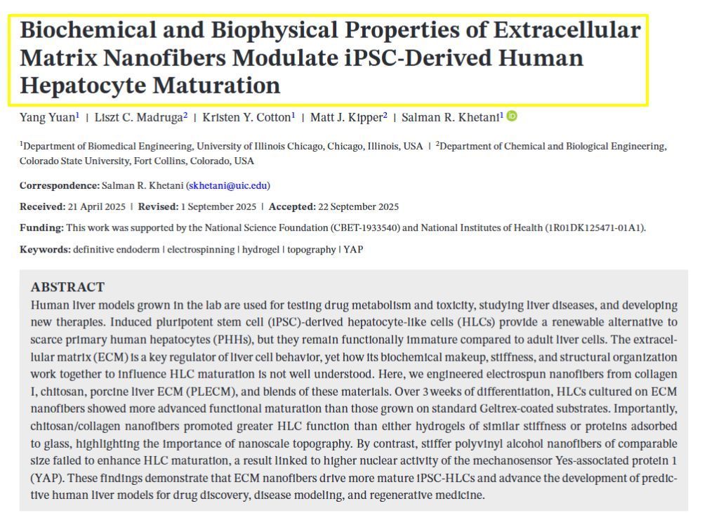
Related MTMLab Publication: https://pubmed.ncbi.nlm.nih.gov/39082962/

May 7, 2025
Our latest study addresses a critical challenge in liver tissue engineering: stem cell-derived liver cells (iHeps) typically remain functionally immature, limiting their usefulness for drug testing and disease modeling. Our research team created 3D microtissues using droplet microfluidics technology by: • Encapsulating iHeps in tiny collagen gel droplets (~250 μm diameter) • Coating these structures with various non-parenchymal cells (NPCs) • Testing different combinations and sequences of supporting cells Key findings: 1) Embryonic fibroblasts and liver sinusoidal endothelial cells (LSECs) produced the most mature iHeps compared to other tested cell types 2) Sequential application of cell signals (embryonic fibroblasts first, then LSECs) yielded optimal maturation 3) Specific growth factors like stromal-derived factor-1 alpha were identified as important maturation enhancers 4) Gene expression analysis confirmed that LSEC/iHep microtissues closely resembled adult human liver cells This platform enables researchers to identify critical cellular interactions and molecular signals that drive liver cell maturation, providing valuable insights for developing more physiologically relevant liver models for drug screening and regenerative medicine applications. https://www.sciencedirect.com/science/article/pii/S174270612500193X SIMPLE SUMMARY: Embryonic fibroblasts and liver sinusoidal endothelial cells dramatically improved iHep maturation compared to other cell types tested, producing more functionally mature liver cells. Sequential application proved crucial—adding embryonic fibroblasts first, followed by endothelial cells, yielded optimal maturation. Specific growth factors including stromal-derived factor-1 enhanced this process. This research enables creation of more authentic mini-liver tissues that function like human liver. These improved models support better drug testing, disease research, and regenerative medicine applications.
December 13, 2024
1. Three-Dimensional (3D) Cell Culture Techniques : New 3D cell culture methods have significantly improved the properties of stem cells, enhancing their viability and functionality for tissue regeneration. These techniques allow for more accurate modeling of tissue architecture and function. 2. Engineered Stem Cells : Advances in bioengineering have led to the development of "engineered stem cells," which are modified to enhance their regenerative capabilities. These next-generation stem cells are designed to be more effective in tissue repair and regeneration. 3. Injectable Biomimetic Hydrogels : Researchers have developed advanced injectable hydrogels that mimic natural tissue environments. These hydrogels hold significant promise for tissue engineering applications, providing a supportive matrix for stem cell growth and differentiation. 4. Integration with Tissue Scaffolds : There have been significant improvements in integrating stem cells with biomaterial scaffolds. These scaffolds provide structural support and enhance the differentiation and growth of stem cells into specific tissue types, improving the outcomes of regenerative treatments. 5. Gene Editing and mRNA Technologies : Techniques like CRISPR and mRNA-based therapies are being used to modify stem cells at the genetic level, enhancing their ability to regenerate tissues. These technologies allow for precise control over stem cell behavior and function.
December 13, 2024
The MTM lab has experienced considerable growth over the last several years at the University of Illinois Chicago!
More Posts