Understanding Histological Patterns

Histological patterns provide crucial insights into the intricate architecture of various bodily tissues such as muscle, bone marrow, connective tissue, blood, and nerve fibers. The process starts with meticulous tissue preparation, where samples are carefully collected and preserved to retain structural details.


Staining is a pivotal step in histology, enhancing contrast in the microscopic images. For instance, hematoxylin and eosin stain distinguishes cellular components, making nuclei appear blue-purple and cytoplasm pink. Specialized stains like Giemsa highlight mast cells, which play a role in immune response and allergic reactions.


When viewing muscle tissue, characteristic striations and banding patterns are apparent, reflecting the organized arrangement of myofibrils. Observing bone marrow reveals a complex milieu of hematopoietic cells involved in blood cell formation. In connective tissue, an extracellular matrix embedded with fibroblasts and collagen fibers becomes visible, providing the framework for tissue support and elasticity.


The histological examination of blood can demonstrate the abundance and ratio of the various cell types, essential for diagnosing disorders. Nerve tissue analysis shows a network of neurons and supporting glial cells, crucial for understanding neurological function and pathology.


Recognition of these patterns is fundamental for correlating structural organization with physiological function and identifying both normal and pathological conditions. The histologic study is thus indispensable for medical research and clinical diagnosis.

May 7, 2025
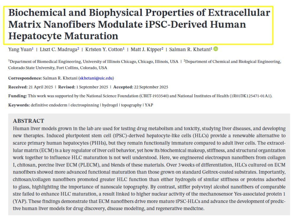
Our latest study addresses a critical challenge in liver tissue engineering: stem cell-derived liver cells (iHeps) typically remain functionally immature, limiting their usefulness for drug testing and disease modeling. Our research team created 3D microtissues using droplet microfluidics technology by: • Encapsulating iHeps in tiny collagen gel droplets (~250 μm diameter) • Coating these structures with various non-parenchymal cells (NPCs) • Testing different combinations and sequences of supporting cells Key findings: 1) Embryonic fibroblasts and liver sinusoidal endothelial cells (LSECs) produced the most mature iHeps compared to other tested cell types 2) Sequential application of cell signals (embryonic fibroblasts first, then LSECs) yielded optimal maturation 3) Specific growth factors like stromal-derived factor-1 alpha were identified as important maturation enhancers 4) Gene expression analysis confirmed that LSEC/iHep microtissues closely resembled adult human liver cells This platform enables researchers to identify critical cellular interactions and molecular signals that drive liver cell maturation, providing valuable insights for developing more physiologically relevant liver models for drug screening and regenerative medicine applications. https://www.sciencedirect.com/science/article/pii/S174270612500193X SIMPLE SUMMARY: Embryonic fibroblasts and liver sinusoidal endothelial cells dramatically improved iHep maturation compared to other cell types tested, producing more functionally mature liver cells. Sequential application proved crucial—adding embryonic fibroblasts first, followed by endothelial cells, yielded optimal maturation. Specific growth factors including stromal-derived factor-1 enhanced this process. This research enables creation of more authentic mini-liver tissues that function like human liver. These improved models support better drug testing, disease research, and regenerative medicine applications.
December 13, 2024
1. Three-Dimensional (3D) Cell Culture Techniques : New 3D cell culture methods have significantly improved the properties of stem cells, enhancing their viability and functionality for tissue regeneration. These techniques allow for more accurate modeling of tissue architecture and function. 2. Engineered Stem Cells : Advances in bioengineering have led to the development of "engineered stem cells," which are modified to enhance their regenerative capabilities. These next-generation stem cells are designed to be more effective in tissue repair and regeneration. 3. Injectable Biomimetic Hydrogels : Researchers have developed advanced injectable hydrogels that mimic natural tissue environments. These hydrogels hold significant promise for tissue engineering applications, providing a supportive matrix for stem cell growth and differentiation. 4. Integration with Tissue Scaffolds : There have been significant improvements in integrating stem cells with biomaterial scaffolds. These scaffolds provide structural support and enhance the differentiation and growth of stem cells into specific tissue types, improving the outcomes of regenerative treatments. 5. Gene Editing and mRNA Technologies : Techniques like CRISPR and mRNA-based therapies are being used to modify stem cells at the genetic level, enhancing their ability to regenerate tissues. These technologies allow for precise control over stem cell behavior and function.
December 13, 2024
The MTM lab has experienced considerable growth over the last several years at the University of Illinois Chicago!
More Posts