Basics of Histology: Microscopy Techniques, Staining Methods & Tissue Preparation

Histology, regarded as an essential tool in biological and medical sciences, allows the detailed examination of tissues and cells. This section provides an overview of the foundational techniques used to study the microscopic structures of biological specimens.


Microscopy Techniques


Light microscopy and electron microscopy are pivotal in histology. Light microscopy, suitable for most routine investigations, uses visible light to illuminate specimens for magnification up to around 1000 times. Alternatively, electron microscopy employs a beam of electrons, greatly surpassing light microscopy in resolution and magnification, which can reveal ultrastructural details.


Staining Methods


The process of staining is crucial for distinguishing and studying the morphological features and chemical components of biological tissues. Common staining methods include hematoxylin and eosin (H&E) for general morphology and various histochemical procedures to identify specific elements within cells and tissues. These techniques provide insight into the anatomy and physiology by visualizing different components uniquely.


Tissue Preparation


Tissue preparation involves multiple steps: fixation, dehydration, sectioning, and embedding. Tissues are initially fixed to preserve structure and prevent decay. Dehydration then follows to remove water, making the tissue amenable to being encased in a solid medium. Thin sections are cut using a microtome, and finally, tissues are embedded in a supportive matrix, facilitating detailed microscopic analysis.

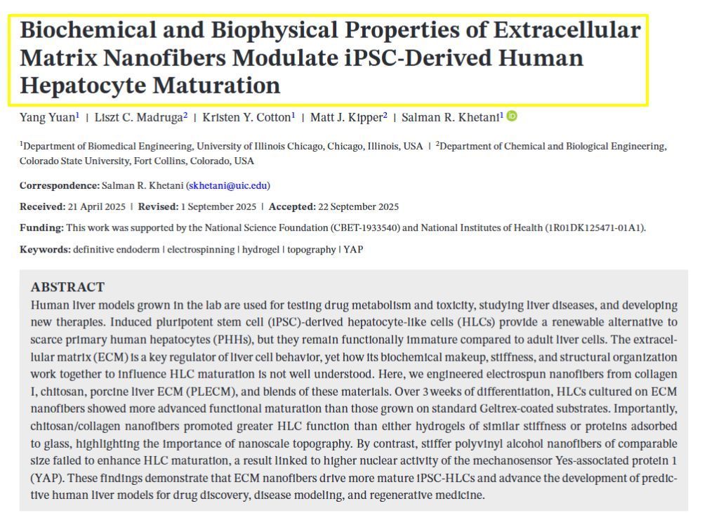
May 7, 2025
Our latest study addresses a critical challenge in liver tissue engineering: stem cell-derived liver cells (iHeps) typically remain functionally immature, limiting their usefulness for drug testing and disease modeling. Our research team created 3D microtissues using droplet microfluidics technology by: • Encapsulating iHeps in tiny collagen gel droplets (~250 μm diameter) • Coating these structures with various non-parenchymal cells (NPCs) • Testing different combinations and sequences of supporting cells Key findings: 1) Embryonic fibroblasts and liver sinusoidal endothelial cells (LSECs) produced the most mature iHeps compared to other tested cell types 2) Sequential application of cell signals (embryonic fibroblasts first, then LSECs) yielded optimal maturation 3) Specific growth factors like stromal-derived factor-1 alpha were identified as important maturation enhancers 4) Gene expression analysis confirmed that LSEC/iHep microtissues closely resembled adult human liver cells This platform enables researchers to identify critical cellular interactions and molecular signals that drive liver cell maturation, providing valuable insights for developing more physiologically relevant liver models for drug screening and regenerative medicine applications. https://www.sciencedirect.com/science/article/pii/S174270612500193X SIMPLE SUMMARY: Embryonic fibroblasts and liver sinusoidal endothelial cells dramatically improved iHep maturation compared to other cell types tested, producing more functionally mature liver cells. Sequential application proved crucial—adding embryonic fibroblasts first, followed by endothelial cells, yielded optimal maturation. Specific growth factors including stromal-derived factor-1 enhanced this process. This research enables creation of more authentic mini-liver tissues that function like human liver. These improved models support better drug testing, disease research, and regenerative medicine applications.
December 13, 2024
1. Three-Dimensional (3D) Cell Culture Techniques : New 3D cell culture methods have significantly improved the properties of stem cells, enhancing their viability and functionality for tissue regeneration. These techniques allow for more accurate modeling of tissue architecture and function. 2. Engineered Stem Cells : Advances in bioengineering have led to the development of "engineered stem cells," which are modified to enhance their regenerative capabilities. These next-generation stem cells are designed to be more effective in tissue repair and regeneration. 3. Injectable Biomimetic Hydrogels : Researchers have developed advanced injectable hydrogels that mimic natural tissue environments. These hydrogels hold significant promise for tissue engineering applications, providing a supportive matrix for stem cell growth and differentiation. 4. Integration with Tissue Scaffolds : There have been significant improvements in integrating stem cells with biomaterial scaffolds. These scaffolds provide structural support and enhance the differentiation and growth of stem cells into specific tissue types, improving the outcomes of regenerative treatments. 5. Gene Editing and mRNA Technologies : Techniques like CRISPR and mRNA-based therapies are being used to modify stem cells at the genetic level, enhancing their ability to regenerate tissues. These technologies allow for precise control over stem cell behavior and function.
December 13, 2024
The MTM lab has experienced considerable growth over the last several years at the University of Illinois Chicago!
More Posts